Ana Cláudia Moura1, Joana Queiros Pereira2, Herminia Choon3, Lorena Stella1, Tiago Milheiro Silva1, Catarina Gouveia1.
1Pediatrics Infectious Diseases Unit, Hospital Dona Estefânia, Unidade Local de Saúde de São José, Lisbon, Portugal,
2Paediatric Surgery Unit, Hospital Dona Estefânia, Unidade Local de Saúde de São José, Lisbon, Portugal,
3Microbiology Unit, Hospital Dona Estefânia, Unidade Local de Saúde de São José, Lisbon, Portugal.
ADDRESS FOR CORRESPONDENCE
Ana Cláudia Moura, Hospital Dona Estefânia, Rua Jacinta Marto 8ª 1169-045, Lisboa, Portugal.
Email: anaclaudiavazdemoura@gmail.com | | Abstract | | Rib tuberculosis is a rare form of tuberculosis. We report the case of an immunocompetent adolescent, with a thoracic cold abscess, presenting with a unilateral painless pectoral mass. Imaging revealed a lytic rib lesion with a perilesional large abscess. Biopsy allowed isolation of Mycobacterium tuberculosis resistant to pyrazinamide. Early diagnosis and prompt treatment are critical to prevent complications. | | | | Keywords | | Cold abscess, Tuberculosis, Chest wall, Mycobacterium tuberculosis. | | | | Introduction | | Musculoskeletal tuberculosis (TB) is a rare form of extrapulmonary TB, with spinal TB being the most frequent.1,2,3 Rib involvement is uncommon, comprising 1-5% of musculoskeletal TB cases and 0.1% of total TB cases.1 Although it can involve any part of the rib, the anterior chest wall is the most frequently affected area.3 Rib TB is twice as common in males, primarily affecting individuals aged 15 to 30 years.4 Fewer than 50% of patients have concurrent active pulmonary TB.1 The clinical presentation of rib TB typically includes a painful or non-tender chest wall mass or chest pain, with variable consistency.1,3 It typically appears as a swelling without signs of inflammation, commonly referred to as a cold abscess.1,5 In this report, the authors present a case of TB of the rib presenting as a cold abscess of the chest wall in an immunocompetent patient. | | | | Case Report | A 17-year-old male, originally from Bangladesh and residing in Portugal for two years, presented with a left retro-mammary swelling for one month, progressively increasing in size. He denied respiratory and systemic symptoms such as fever, weight loss, and night sweats. Unknown status of BCG vaccination. The patient had no significant past medical history, but his 15-year-old sister was recently hospitalized for suspected (later confirmed) tuberculous cervical lymphadenitis. Additionally, his mother, undergoing anti-tumor necrosis factor therapy, had latent TB and completed a three-month treatment course, six months earlier.
On physical examination he looked well, a firm, non-fluctuant, left retro-mammary swelling, measuring 8x6 cm, was observed. No tenderness, local rise of temperature or redness of the overlying skin was present. Pulmonary auscultation was normal.
Blood tests showed elevated C-reactive protein (37 mg/L, NR<5 mg/L) and erythrocyte sedimentation rate (32 mm/h, NR<20 mm/h). HIV infection was excluded. IGRA QuantiFERON was positive. Mantoux test was also positive (30x20 mm reaction with an associated ulcer).
Figure 1a. Axial CT section showing a hypodense lesion, with a lytic destruction of the anterior arc of the left 3rd rib, causing cortical irregularity (red arrow) and infiltration of the left pectoralis minor muscle; Figure 1b. Purulent content aspirated from the thoracic mass.
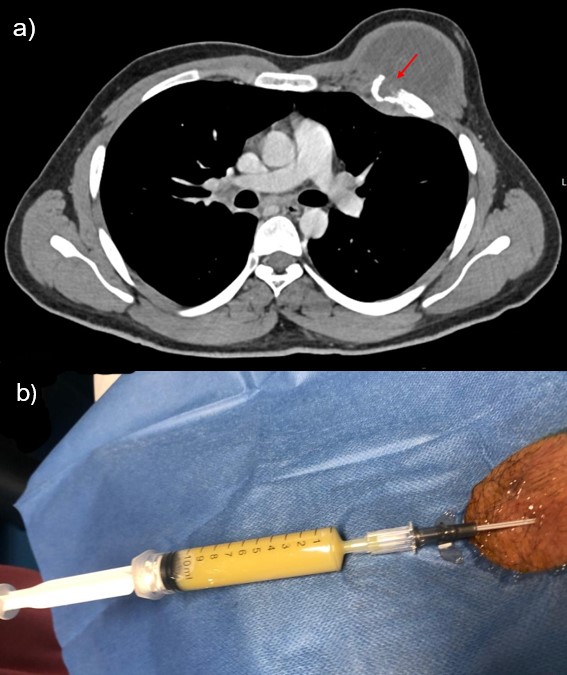
Thoracic ultrasound, X-ray and tomography revealed a large necrotic collection measuring 84x65x61 mm (LxAPxT) in the left pectoralis minor muscle, with expansion and lytic destruction of the anterior segment of the left 3rd rib, causing cortical irregularity (Figure 1a). Several enlarged left axillary lymph nodes were identified, the largest measuring 12x9 mm. No other abnormalities were observed in the pulmonary X-ray and abdominal ultrasound. A fine-needle aspiration of the thoracic abscess was performed under ultrasound guidance, yielding approximately 30 cc of yellow purulent fluid (Figure 1b). Samples from the aspirate were sent for culture and Nucleic Acid Amplification Test (NAAT) for M. tuberculosis. Given the high TB suspicion, the patient was started on anti-tuberculosis treatment (ATT) consisting of daily isoniazid 300 mg, rifampicin 600 mg, pyrazinamide 1500 mg and ethambutol 1200 mg, complemented with pyridoxine 40 mg, under a directly observed therapy regimen. Mycobacterium tuberculosis complex was isolated from the aspirate (NAAT was also positive), and the strain was found to be sensitive to isoniazid, ethambutol, and rifampicin, but resistant to pyrazinamide. pyrazinamide was discontinued, while the remaining medications were continued. ethambutol was stopped after 2 months of treatment. The patient has completed 12 months of ATT. A favorable clinical response has been observed, with a significant reduction in the mass size during follow-up visits. | | | | Discussion | Rib TB can pose significant diagnostic challenges, especially when presenting as a thoracic abscess.2,5,6 In this case, the patient presented with a cold abscess, a typical pattern of musculoskeletal TB, and distinct from pyogenic infections. TB was strongly suspected due to patient's background from a TB-endemic country and the family TB history.2,6 Unfortunately, the patient, and his sister, were not screened for TB upon his mother’s diagnosis of latent TB, representing a missed opportunity for early detection and intervention.
Chest computed tomography is regarded as the imaging modality of choice for assessing TB chest wall lesions.1,3,4,5 It effectively reveals bone erosion, the characteristics and extent of soft tissue collections, associated intrathoracic lesions and lymphadenopathy.3,5 In the presence of lytic lesions, rib widening, periosteal reaction and/or sequestrum TB should be considered.3,7 TB is the second most common cause of osteolytic rib lesions, after neoplasms.7 Other infections associated with rib destruction include typhoid and paratyphoid fever, actinomycosis, syphilis, blastomycosis, and brucellosis.1,7,8 Ultrasound can show the internal architecture of the mass and assess muscle and bone involvement.3,4,5 The chest radiograph is the initial test.3,7 In this case, imaging revealed a well-defined necrotic collection involving the pectoralis minor muscle, with associated bone destruction and axillary lymphadenopathy, but no pulmonary or mediastinal involvement. These findings are not pathognomonic of TB. The cultural isolation of Mycobacterium spp. remains essential for definitive diagnosis and appropriate treatment.1,3,8 Specimens can be obtained through fine-needle aspiration cytology (FNAC) in the presence of a thoracic abscess, as illustrated in our case, obviating the need of an open biopsy.1,3,5,8 FNAC is a minimally invasive first-line diagnostic approach, particularly when guided by imaging.3,7,8 It may demonstrate cytological features suggestive of TB, such as granulomatous inflammation with caseous necrosis.3,8 However, its sensitivity is limited by sample adequacy.1,3,6,8 The decision between FNAC and open biopsy should balance diagnostic sensitivity with procedural risk, based on clinical, imaging, and expertise factors.3,8
There is no standardized therapeutic protocol for rib TB.4 Medical treatment follows the same first-line regimen as pulmonary TB, including isoniazid, rifampicin, ethambutol, and pyrazinamide (PZA), with the exception of treatment duration. In osteoarticular TB, treatment should be extended to 12 months, with a two-month intensive phase followed by a ten-month continuation phase.9 Isolated PZA resistance is uncommon but requires modification of therapy, including extension of treatment duration and exclusion of PZA once resistance is confirmed by drug susceptibility testing.10 The regimen should maintain first-line agents such as isoniazid, rifampicin, and ethambutol.10 A later-generation fluoroquinolone, such as levofloxacin or moxifloxacin, may be added in selected cases to enhance bactericidal activity and compensate for the loss of PZA’s sterilizing effect.10 Treatment duration is generally nine to twelve months for pulmonary tuberculosis with isolated PZA resistance and at least twelve months for osteoarticular disease.1,6,9 The pyridoxine supplementation should be initiated in adolescents during isoniazid treatment, to prevent peripheral neuropathy.9 Complete surgical resection is controversial.1,11 Surgical intervention may be needed for extensive tissue damage, draining sinuses, or failure of medical treatment.1,4,11 Our patient was treated by aspiration of the cold abscess and ATT for 12 months, without the need for further surgical intervention. | | | | Conclusion | | This case report emphasizes the need to consider TB in the differential diagnosis of a chest wall mass and the importance of screening household contacts of patients diagnosed with TB. Early diagnosis and treatment are crucial to prevent complications. | | | | Compliance with Ethical Standards | | Funding None | | | | Conflict of Interest None | | |
- Cherry JD, Demmler-Harrison, G. J., Kaplan, S. L., Steinbach, W. J., & Hotez, P. J. (Eds.). In: Feigin and Cherry's Textbook of Pediatric Infectious Diseases. Vol 2. 8th ed.2018:959-971.
- BORGES M, ROCHA AP, DE MACEDO CV, et al. Pediatric Tuberculosis: 12 Years of Experience in a Tertiary Referral Center in Portugal Tuberculose em Idade Pediátrica: Experiência de 12 Anos num Centro Terciário de Referência em Portugal. 2022. [CrossRef] [PubMed]
- Pattamapaspong N, Kanthawang T, Bouaziz MC, Ladeb MF, Hammami N, Peh WC. Imaging of musculoskeletal tuberculosis. British Journal of Radiology. 2024;97(1153):1-12. [CrossRef] [PubMed] [PMC free article]
- Hussein M, Abdelhadi A, Elarabi A, Rashid I, Alabbas A, Aladab A. Extrapulmonary Tuberculosis masquerading as chest wall malignancy: just never ceases to surprise! IDCases. 2021;24:e01114. [CrossRef] [PubMed] [PMC free article]
- Abbassi H, Bannour B, Yacoub S, et al. From Plasmacytoma to Rib Tuberculosis: The Case of A Breast Mass With An Unexpected Diagnosis. European Journal of Breast Health. 2025;21(1):85. [CrossRef] [PubMed] [PMC free article]
- Newton SM, Brent AJ, Anderson S, Whittaker E, Kampmann B. Paediatric tuberculosis. The Lancet infectious diseases. 2008;8(8):498-510. [CrossRef] [PubMed] [PMC free article]
- Saldaña NG, Trujillo DMG, Morales SAS, Corona QV, Jimenez FT, Olguin HJ. Tuberculosis of the ribcage by dissemination of Koch bacilli. The Journal of Infection in Developing Countries. 2022;16(07):1221-1225. [CrossRef] [PubMed]
- Handa U, Garg S, Mohan H, Garg SK. Role of fine‐needle aspiration cytology in tuberculosis of bone. Diagnostic cytopathology. 2010;38(1):1-4. [CrossRef] [PubMed]
- WHO operational handbook on tuberculosis. Module 5: management of tuberculosis in children and adolescents. In. Geneva: World Health Organization; 2022. Licence: CC BY-NC-SA 3.0 IGO.
- Lange C, Dheda K, Chesov D, Mandalakas AM, Udwadia Z, Horsburgh CR, Jr. Management of drug-resistant tuberculosis. Lancet. 2019;394(10202):953-966. [CrossRef] [PubMed] [PMC free article]
- Keum D-Y, Kim J-B, Park C-K. Surgical treatment of a tuberculous abscess of the chest wall. The Korean journal of thoracic and cardiovascular surgery. 2012;45(3):177. [CrossRef] [PubMed] [PMC free article]
|
| Cite this article as: | | Moura A C, Pereira J Q, Choon H, Stella L, Silva T M, Gouveia C. Slow-Growing Unilateral Pectoral Mass: Would You Worry?. Infection Child J. 2026;1. |
|