Endobronchial Tuberculosis - To Operate Or Not To Operate
|
|
Endobronchial Tuberculosis - To Operate Or Not To Operate
08/06/2026
08/06/2026
https://www.pediatriconcall.com/Journal/images/journal_cover.jpg
Dhruvi Shah1, Darsita Jatakia2, Theertha Ramesh1, Ira Shah1.
1Pediatric DR-TB Center of Excellence, Department of Infectious Diseases, Bai Jerbai Wadia Hospital for Children, Acharya Donde Marg, Parel, Mumbai, Maharashtra, India,
2Department of Pediatrics, Seth G S Medical College and KEM Hospital, Acharya Donde Marg, Parel, Mumbai, Maharashtra, India.
ADDRESS FOR CORRESPONDENCE
Darsita Jatakia, Seth G S Medical College and KEM Hospital, Acharya Donde Marg, Parel, Mumbai, Maharashtra 400012, India.
Email: jatakia.darsita@gmail.com
Endobronchial Tuberculosis, Lobectomy, bronchial stenosis
|
Clinical Problem
A 14-year-old girl presented with fever, cough with expectoration and breathing difficulty since 10 days, in May 2024. Sputum showed rifampicin sensitive (RS) Mycobacterium tuberculosis (MTB) on Xpert MTB/Rif Assay. She was started on anti-tubercular therapy (ATT) of isoniazid (H), rifampicin (R), pyrazinamide (Z) and ethambutol (E). In May 2025, she presented with cough and difficulty breathing. Her weight was 39 kg and height was 152cm. Heart rate and blood pressure were normal, respiratory rate was 22/min. Laboratory investigations are depicted in Table 1. On examination, air entry was reduced on right side. Chest X-ray revealed collapse of middle and lower lobe of right lung (Figure 1). Chest Computed Tomography (CT) scan showed irregular narrowing of right lower lobe bronchus, complete occlusion of bronchus intermedius and right lower lobe bronchus, diffuse wall thickening and severe luminal narrowing of right middle lobe bronchus. There was obstructive emphysema of right middle lobe, and collapse consolidation of right lower lobe with necrosis. Fibronodular changes were seen in right upper lobe and in posterobasal segment of left lower lobe. Xpert MTB/Rif Assay of bronchoalveolar lavage detected low RS MTB. Perfusion scan showed total 31% uptake in right lung, with 7% and 1% uptake in right middle and right lower lobe, respectively. Bronchoscopy performed showed thin fibrotic bands around mid-trachea and granulation in anterior wall. Bronchus intermedius showed fibrostenotic type of endobronchial TB (EBTB). Right upper lobe showed mild stenosis with mild secretions. She was admitted for lobectomy which was deferred considering significant inflammation seen on bronchoscopy, distal to right bronchus intermedius, with stenosis. (3.8 Fr bronchoscope could not be negotiated). She was additionally advised levofloxacin and steroids.
Figure 1. Chest Xray showing collapse of the middle and lower lobe of the right lung.
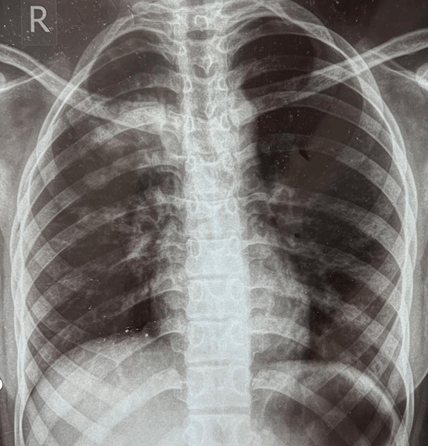
Table 1. Laboratory investigations at presentation.
| Parameter |
Day of presentation |
| Haemoglobin (g/dl) |
11.5 |
| Total White Count (cells/cumm) |
4870 |
| Platelet count (cells/cumm) |
369000 |
| SGOT/SGPT (U/L) |
33/21 |
| Sodium (mEq/l) |
137 |
| Potassium (mEq/l) |
4.8 |
| Chlorine (mEq/L) |
102 |
| Calcium (mEq/L) |
9.7 |
| Blood Urea Nitrogen (mg/dl) |
6 |
| S. Creatinine (mg/dl) |
0.47 |
|
| |
When should patients with endobronchial TB undergo lobectomy?
|
|
|
Discussion
EBTB represents a distinct manifestation of pulmonary tuberculosis (TB), characterized by infection of tracheobronchial tree, with more than 50% cases presenting at <35 years of age. When confirmed with flexible bronchoscope, children, 30-60% cases of uncomplicated primary TB reported endobronchial involvement.1 While EBTB can affect any part of bronchial tree, primary bronchi, bilateral superior bronchi and right middle lobar bronchus are commonly affected. It can be single or multiple level EBTB, based on number of bronchial levels involved.2 It leads to mucosal and submucosal tuberculous infiltration, ulceration, granuloma formation, fibroplasiasis and tracheobronchial stenosis.2 Caseous necrosis occurs in tubercular nodules and mucosal ulcers in later stages.2 This can progress to deep-part ulcer into tracheobronchial wall or lead to inflammatory hyperplastic polyps protruding tracheobronchial lumen, which can develop into fibrous hyperplasia and contractures, and cause tracheobronchial stenosis, in advanced stages.2
EBTB presents with dry cough or with bronchorrhea (70-80%), low grade fever (usually seen in advanced cavitatory disease), hemoptysis (15-40%), chest pain (15-25%), localized wheezing, and decreased breath sounds, if endobronchial lesion causes stenosing effect. Systemic features include anorexia, generalised weakness seen in more than 50% patients.2 More than 90% cases present as complications caused by different degrees of bronchostenosis.1 Primary management of EBTB remains ATT, often supplemented with systemic corticosteroids during active inflammatory phase to reduce mucosal edema and prevent progression to fibrosis.1 Despite appropriate ATT, progressive fibrostenotic disease can develop, causing irreversible bronchial obstruction, lobar collapse, and recurrent infections.3 Bronchoscopic interventions like balloon dilatation, cryotherapy, laser therapy, or stenting may be considered in selected cases with short-segment stenosis or predominantly inflammatory lesions.3,4 However, these approaches are ineffective once dense fibrosis and fixed airway narrowing have developed.3,4
Surgical resection, including lobectomy, is reserved for carefully selected cases including irreversible fibrostenotic airway disease, non-functional lung segments, recurrent or persistent post-obstructive infections, massive hemoptysis, or failure of medical and bronchoscopic management.3,5 Preoperative assessment should demonstrate minimal contribution of affected lung segment to overall pulmonary function, typically confirmed by perfusion scintigraphy or functional imaging.5
In our case, patient had completed ATT but developed progressive respiratory symptoms with radiological evidence of middle and lower lobe collapse. Bronchoscopy revealed fibrostenotic EBTB involving bronchus intermedius, with inability to negotiate even a small-caliber bronchoscope, indicating advanced irreversible disease. Perfusion scan demonstrated markedly reduced functional contribution from right middle and lower lobes (7% and 1%, respectively), fulfilling functional criteria for surgical consideration.
Timing of surgery is critical in EBTB, surgery is ideally deferred until active inflammatory phase has subsided, as operating during active disease increases risk of postoperative complications and restenosis.5 In our patient, lobectomy was appropriately deferred due to significant distal inflammation seen on bronchoscopy, and escalation of medical therapy with corticosteroids and levofloxacin was undertaken to control inflammation prior to reconsidering surgery.
This case shows that lobectomy in EBTB is not dictated by microbiological status alone, but by structural and functional sequelae of disease. Early recognition of EBTB, close bronchoscopic follow-up, and timely referral for surgical evaluation are essential to prevent long-term morbidity.3
|
| |
| Compliance with ethical standards |
|
Funding: None
|
|
|
Conflict of Interest: None
|
- Dey A, Shah I. Infantile endobronchial tuberculosis. J Family Med Prim Care. 2019 Jan;8(1):299-301. [CrossRef] [PubMed] [PMC free article]
- Shahzad T, Irfan M. Endobronchial tuberculosis-a review. J Thorac Dis. 2016 Dec;8(12):3797-3802. [CrossRef] [PubMed] [PMC free article]
- Thiet TT, Nguyen TV, Truong TH, et al. Long-term outcomes of surgical reconstruction for post-tuberculosis tracheobronchial stenosis: a 7-year follow-up study in a tuberculosis-endemic region. J Thorac Dis. 2024;16(3):1021-1031. [CrossRef] [PubMed] [PMC free article]
- Mohd Esa NY, Lee YF, Chong VH, et al. Bronchoscopic features and morphology of endobronchial tuberculosis. J Clin Med. 2022;11(3):676. [CrossRef] [PubMed] [PMC free article]
- Pulle MV, Asaf BB, Puri HV, Bishnoi S, Kumar A. Surgical intervention is safe, feasible, and effective in tubercular tracheobronchial stenosis. Lung India. 2021;38(6):541-546. [CrossRef] [PubMed] [PMC free article]
|
|
| |
|
DOI:
|
| |
Cite this article as:
Shah D, Jatakia D, Ramesh T, Shah I. Endobronchial Tuberculosis - To Operate Or Not To Operate. Infection Child J. 2026;1.
|