|
Recognising and Managing Ocular Toxoplasmosis in Children: A Case Report
Mariana Reis1, Joana Capela1, Inês Almeida1, Ana Teresa Martins2, Sara Geitoeira1, Luísa Gaspar3, Andreia Fernandes1.
1Department of Pediatrics, Faro Hospital, ULS Algarve, Faro, Portugal,
2Department of Ophtalmology, Faro Hospital, ULS Algarve, Faro, Portugal,
3Department of Pediatric and Neonatology Intensive Medicine, ULS Algarve, Faro, Portugal.
ADDRESS FOR CORRESPONDENCE
Mariana Freitas Reis, Rua Leão Penedo, Faro.
Email: mari.reis@sapo.pt
Ocular toxoplasmosis, Chorioretinitis lesion, Toxoplasma gondii
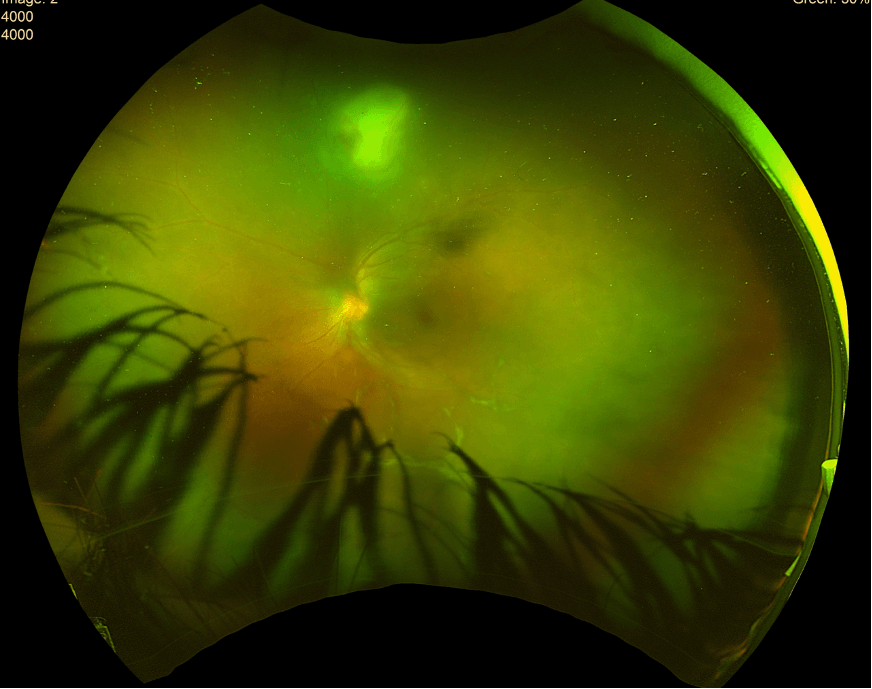
A previously healthy 12-year-old boy from Brazil, living in Portugal for the past two years, with no relevant antenatal, perinatal or past medical history, presented with a two-month history of mild conjunctival hyperaemia, decreased visual acuity and intermittent left ocular pain. Ophthalmological examination revealed reduced visual acuity in the left eye (1/10), inferior keratic precipitates, peripheral vitritis and a solitary superior white chorioretinitis lesion (Figure 1).
The patient denied systemic symptoms, including fever, asthenia, weight loss, cough, night sweats, rash, arthralgia, arthritis, diarrhoea, or oral ulcers. He had occasional contact with dogs and cats at a local football pitch, but denied exposure to other animals. There was no recent international travel.
Figure 1. Ultra-widefield pseudo-colour retinography of the left eye showing a white focal retinitis with overlying vitreous inflammation (headlight-in-fog sign), along with papillitis and widespread vasculitis. The right eye showed no abnormalities.
|
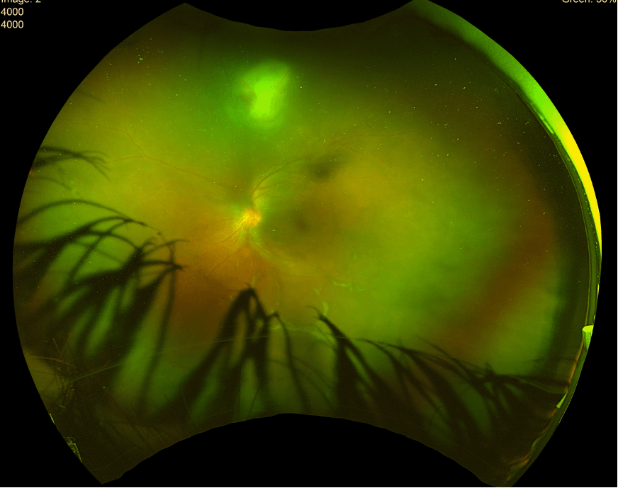
Figure 1. Ultra-widefield pseudo-colour retinography of the left eye showing a white focal retinitis with overlying vitreous inflammation (headlight-in-fog sign), along with papillitis and widespread vasculitis. The right eye showed no abnormalities.
|
What’s the diagnosis?
The eye findings were characteristic of ocular toxoplasmosis (OT). Laboratory evaluation revealed positive Toxoplasma gondii IgG, with negative IgM. Inflammatory parameters were negative, liver enzymes were mildly elevated (ALT 56 U/L, AST 36 U/L), angiotensin-converting enzyme was increased (129 U/L). Additional autoimmune testing (including complement 3 and 4, rheumatoid factors, anti-dsDNA, ANA, ANCA-PR3, ANCA-MPO and glucose-6-phosphate dehydrogenase) and infectious serologies (for Borrelia burgdorferi, Brucella, Treponema pallidum, Bartonella henselae, Mycobacterium tuberculosis, Toxocara canis, hepatitis B, HIV, Epstein–Barr virus, herpes simplex virus, varicella–zoster virus and cytomegalovirus) were all negative.
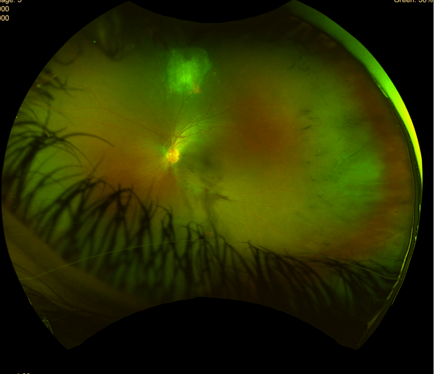
Despite serology not indicating acute infection, clinical suspicion for OT remained high, and treatment with pyrimethamine, sulfadiazine and folinic acid was initiated, along with topical corticosteroids, bromfenac and cyclopentolate. However, within two days he developed haematuria and acute kidney injury, attributed to sulfadiazine-induced crystalluria. This prompted a switch to systemic azithromycin. Following this adjustment, the patient demonstrated progressive clinical improvement (Figure 2).
Figure 2. Follow-up after 2 months of treatment: imaging demonstrates improved lesion definition with pigmentary alteration and reduced surrounding retinal haze, consistent with lesion organization and scarring.
OT is the most common cause of infectious posterior uveitis in children and young adults. Diagnosis in immunocompetent patients is primarily clinical, based on unilateral focal necrotising chorioretinitis with vitritis, findings that were present in this patient.1 Positive IgG and negative IgM serology indicated prior exposure rather than recent infection, supporting reactivation of latent toxoplasmosis, a common mechanism of OT. Although cat exposure was limited, infection may occur through contact with contaminated soil, water or food, and the patient’s origin from Brazil, a highly endemic region where more virulent strains circulate, may have increased his risk of prior infection4. Because the presentation was typical and treatment response was favourable, ocular fluid PCR or intraocular antibody testing was not required.2
Early treatment is essential, as untreated disease may progress over weeks to months and result in retinal scarring, cataract, glaucoma, retinal detachment and permanent visual impairment.3 In this case, reduced visual acuity and vitritis were present, but no irreversible complications developed. Standard therapy includes pyrimethamine, sulfadiazine and folinic acid; however, sulfadiazine-induced crystalluria necessitated a switch to azithromycin, with subsequent improvement. Intravitreal clindamycin plus dexamethasone may be considered in cases of systemic intolerance or sight-threatening lesions but was unnecessary in this patient because of the favourable response to systemic therapy.1 |
| |
| Compliance with ethical standards |
|
Funding: None
|
|
|
Conflict of Interest: None
|
|
- Casado FC, Samper AD. Sulfonamide allergy and alternative treatments in ocular toxoplasmosis. Rom J Ophthalmol. 2025;69(2):147-157. doi:10.22336/rjo.2025.25. [CrossRef] [PubMed] [PMC free article]
- Miyagaki M, Zong Y, Yang M, Zhang J, Zou Y, Ohno-Matsui K, et al. Ocular toxoplasmosis: advances in Toxoplasma gondii biology, clinical manifestations, diagnostics, and therapy. Pathogens. 2024;13: DOI:10.3390/pathogens13100898 [CrossRef] [PubMed] [PMC free article]
- Furtado JM, Smith JR. Pediatric uveitis. J Fam Med Prim Care. 2024;13(11):4815-4818. doi:10.4103/jfmpc.jfmpc_923_24. PMID:39722929; PMCID:PMC11668373. [CrossRef] [PubMed] [PMC free article]
- De-La-Torre A, Mejía-Salgado G, Eraghi AT, Pleyer U. Age and ocular toxoplasmosis: a narrative review. FEMS Microbes. 2025;6. DOI: 10.1093/femsmc/xtaf002 [CrossRef] [PubMed] [PMC free article]
|
|
| |
Cite this article as:
Reis M, Capela J, Almeida I, Martins A T, Geitoeira S, Gaspar L, Fernandes A. Recognising and Managing Ocular Toxoplasmosis in Children: A Case Report. Infection Child J. 2026;1.
|