Zahabiya Nalwalla, Tsering Yangchen Dirkhipa, Ira Shah.
Department of Pediatric Infectious Diseases, B J Wadia Hospital for Children, Mumbai, India.
ADDRESS FOR CORRESPONDENCE
Tsering Yangchen Dirkhipa, Department of Pediatric Infectious Diseases, B J Wadia Hospital for Children, Mumbai, India.
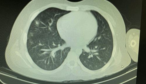
Email: tseringyangchen000@gmail.com | | Abstract | | Nocardiosis is an uncommon infection with an increased susceptibility in immunocompromised individuals. Central venous catheter (CVC) associated nocardiosis has been reported in both adults and children but peripherally inserted central catheter (PICC) associated disseminated nocardiosis has not been reported previously. We present a child with Ewing’s sarcoma with PICC associated disseminated nocardiosis. | | | | Keywords | | Central Catheter, PICC, Disseminated Nocardiosis, Ewings Sarcoma. | | | | Introduction | | Nocardia is a ubiquitous, gram positive, weakly acid-fast filamentous bacilli causing fatal disease in immunocompromised individuals.1 Although central venous catheter (CVC) associated nocardiosis has been seen in both adults and children1,2 and peripherally inserted central catheter (PICC) associated nocardiosis has been seen in adults3, we present a case of PICC linked disseminated nocardiosis in a child with Ewing’s sarcoma undergoing chemotherapy, which has not been reported previously in children. | | | | Case Report | A 12 year old boy was diagnosed with Non-metastatic Ewing’s sarcoma of the left scapula in July 2021. He was started on chemotherapy and a PICC line was inserted for the same at the initiation of chemotherapy. He underwent left scapular resection in December 2021. In May 2022, he presented to us with fever for three days and cough with expectoration for 2 days. In May 2022, he was on 6-mercaptopurine, prednisolone and cotrimoxazole prophylaxis. (5 mg/kg once a week). On examination, his weight was 55.3 kg (between 90th and 97th centile) and height was 158.2 cm (between 75th and 90th centile). His vital parameters were normal. On auscultation of the chest, crepitations were heard in the inframammary and infrascapular areas bilaterally. Other systemic examinations were normal. His hemoglobin was 9.1 g/dl, total leukocyte count was 10,6100 cells/cumm, platelet count was 439,000 cells/cumm and CRP was 13.6 mg/dl. Urine and stool examination were normal. PICC line and peripheral blood culture grew Nocardia species resistant to amoxicillin-clavulanic acid and ciprofloxacin and sensitive to cotrimoxazole and linezolid. Chest X-ray showed bilateral lower zone non-homogenous opacities and CT chest showed multiple nodules in bilateral lung parenchyma with a consolidatory patch in the right lower lobe measuring 3.1x1.9 cm. (Figure 1). MRI brain was normal. He was started on injection meropenem (40 mg/kg/dose 8 hourly) and linezolid (10 mg/kg/dose 8 hourly) for 2 weeks and cotrimoxazole was increased to (20 mg/kg/day) for 6 months. 6 MP and prednisolone was continued
PICC line was removed. Repeat blood culture after 4 weeks did not grow any organism. A repeat CT chest after 3 months of therapy showed resolution of nodules in the lung. On his last follow up, in December 2022, he was clinically well.
Figure 1. CT Chest showing multiple nodules in bilateral lung parenchyma with a consolidation in the right lower lobe measuring 3.1x1.9 cm.
 | | | | Discussion | Nocardiosis is an infection caused by gram positive weakly acid-fast positive filamentous bacillus Nocardia spp.1 The infection as such is uncommon and individuals with an incompetent immune system are the ones susceptible to this ubiquitous organism.2 Risk factors include poorly controlled HIV infection, patients on long-term corticosteroid treatment, solid organ and hematopoietic stem cell transplant recipients, individuals with malignancy and primary immunodeficiency especially Chronic Granulomatous Disease (CGD).2,4,5 Among these, corticosteroid therapy is considered the most important risk factor for development of nocardiosis.6 The clinical spectrum varies from cutaneous, pleuropulmonary to disseminated disease.4 Primary cutaneous results from direct skin inoculation while pleuropulmonary can occur following inhalation of contaminated aerosolized droplets. Either of these can lead to disseminated nocardiosis.4
Primary cutaneous disease presents in the form of abscesses or tender nodules at the site of inoculation while fever, cough, dyspnea or chest pain can be the presenting complaints in pulmonary nocardiosis.4 When abscesses are formed in two or more locations, the disease is considered disseminated with the symptoms based on the area affected.4,7 Disseminated disease has a relatively higher mortality than the former two.2,8 In cancer patients, central venous catheter and PICC lines are two modalities of delivering chemotherapy.1,9 The child in our case had malignancy (Ewing’s sarcoma) and an indwelling PICC for his chemotherapy, thus both increasing his susceptibility to nocardiosis.2,4
In a study by Al Akhrass et al, they found that nocardia bacteremia was associated with heavy biofilm formation around the CVC.1 This mechanism may be true for PICC as well since both are indwelling catheters used for long duration. There are multiple conditions which may present similarly such as bacterial pneumonia, tuberculosis, parapneumonic pleural effusion, glioblastoma multiforme and sporotrichosis.2,4 Due to the ubiquitous nature of the organism and rarity of the infection, diagnosis becomes challenging. Hence, high index of suspicion is required. Initial evaluation should include bacterial culture of the suspected sites and if dissemination or pulmonary infection is suspected, blood culture should be included as well.4 Chest X-ray and CT scan of chest should be done in case of pulmonary infection along with CT or MRI Brain to rule out CNS dissemination.4
The treatment consists of a minimum of 6 months of antibiotic therapy which should then extend for at least 1 month following resolution of symptoms.4 In immunocompetent individuals, 6-12 months of treatment is often enough while it may be longer in case of immunocompromised individuals.10 Nocardia is susceptible to trimethoprim/sulfamethoxazole (TMP-SMX), amikacin, meropenem, third-generation cephalosporins, minocycline, linezolid, amoxicillin-clavulanic acid, dapsone, moxifloxacin and clarithromycin.4 tmp-smx is the most commonly used first line agent, however the treatment should be individualized and based on culture sensitivity. Monotherapy is sufficient for cutaneous disease in an immunocompetent individual while combination regimen is preferred in case of pulmonary and disseminated disease.4 Prompt removal of catheter with antibiotic initiation is found to be effective in case of indwelling-catheter associated Nocardiosis, as was seen in our patient.1,2 It is advisable to continue monitoring for up to 1 year after antibiotic cessation to detect any relapse.4 | | | | Conclusion | | Nocardiosis creates a diagnostic challenge due to the ubiquitous nature of the organism and its low incidence, hence demanding a high index of suspicion. It should be considered as a differential when an immunocompromised individual with an indwelling catheter presents with suspected symptoms. Treatment includes catheter removal with quick initiation of antibiotic therapy. | | | | Compliance with Ethical Standards | | Funding None | | | | Conflict of Interest None | | |
- Al Akhrass F, Hachem R, Mohamed JA, Tarrand J, Kontoyiannis DP, Chandra J, et al. Central venous catheter-associated Nocardia bacteremia in cancer patients. Emerg Infect Dis. 2011;17:1651-1658. [CrossRef] [PubMed] [PMC free article]
- Watson MEJ, Estabrook MM, Burnham CAD. Catheter-associated Nocardia higoensis bacteremia in a child with acute lymphocytic leukemia. J Clin Microbiol. 2011;49:469. [CrossRef] [PubMed] [PMC free article]
- Njie AB, Mitchell M, Pukkila-Worley R. Peripherally Inserted Central Catheter-Associated Nocardia nova Endocarditis in a Patient Receiving Intravenous Antibiotics for Chronic Lyme Disease. Open Forum Infect Dis. 2021;8:ofab041. doi: 10.1093/ofid/ofab041. [CrossRef] [PubMed] [PMC free article]
- Rawat D, Rajasurya V, Chakraborty RK, Sharma S. Nocardiosis. In: StatPearls [Internet]. Treasure Island (FL): StatPearls Publishing; 2023 Jan. Available from: https://www.ncbi.nlm.nih.gov/books/NBK526075/. [Updated 2022 Aug 1].
- Restrepo A, Clark NM. Infectious Diseases Community of Practice of the American society of Transplantation. Nocardia infections in solid organ transplantation: guidelines from the Infectious Diseases Community of Practice of the American Society of Transplantation. Clin Transplant 2019;33:e13509. [CrossRef] [PubMed]
- Leite IM, Trigueiros F, Martins AM, Fonseca M, Marques T. Disseminated Nocardiosis: A Case Report. Cureus. 2019;11:e5294. [CrossRef]
- Tomas RM, Villanueva RM, Calzada SR, Durantez MS, Tarazona JMV, Alapont MM, Serrano MG. Pulmonary nocardiosis: risk factors and outcomes. Respirology. 2007;12:394-400. [CrossRef] [PubMed]
- Williams E, Jenney AW, Spelman DW. Nocardia bacteremia: A single-center retrospective review and a systematic review of the literature. Int J Infect Dis. 2020;92:197-207. [CrossRef] [PubMed]
- Sapkota S, Sannur R, Naik R. Analysis of Peripherally Inserted Central Catheter Line in Cancer Patients: A Single-Center Experience. South Asian J Cancer. 2020;9:253-256. [CrossRef] [PubMed]
- Aggarwal D, Garg K, Chander J, Saini V, Janmeja AK. Pulmonary nocardiosis revisited: A case series. Lung India. 2015;32:165-168. [CrossRef] [PubMed] [PMC free article]
|
| Cite this article as: | | Nalwalla Z, Dirkhipa T Y, Shah I. Peripherally Inserted Central Catheter (PICC) Associated Disseminated Nocardiosis in a Child with Ewings Sarcoma - A Case Report and Review. Infection Child J. 2026;1. |
|